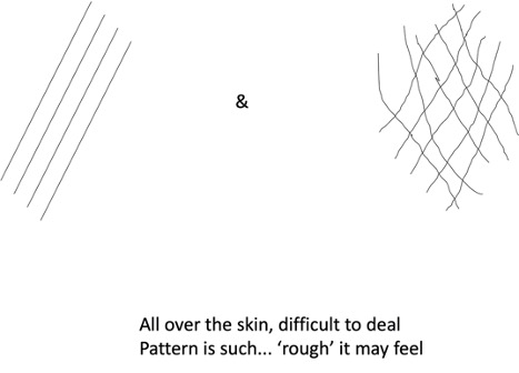
Mid-Month Mindbender
May 21, 2023
Suman Patra
Clue 1
Bumps skin deep; high spikes it stings
Good store in ‘press’; Wow! it’s ‘stress’
In our world though, not always ‘bad’ as you call
Gobbles up whole, spits blood in the mix
Low in ‘numbers’, troubles yet to fix.
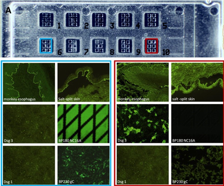
Clue 2
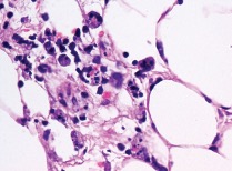 Click on the image to enlarge a bit
What is your diagnosis?
Click here for the answer
April 26, 2023
Suman Patra
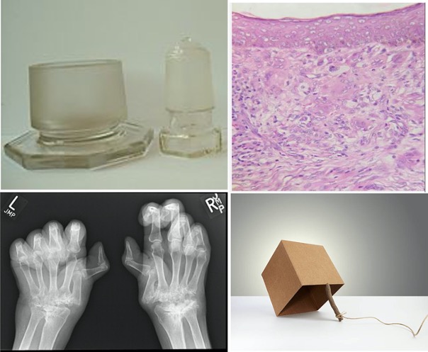 Click on the image to enlarge
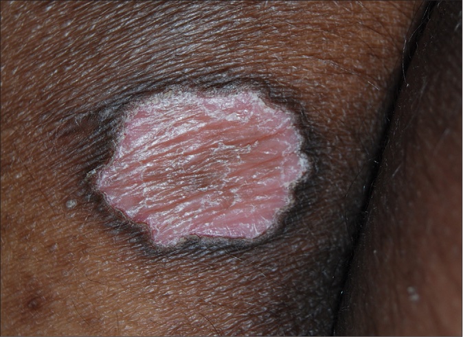
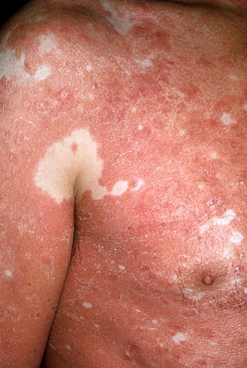
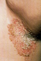
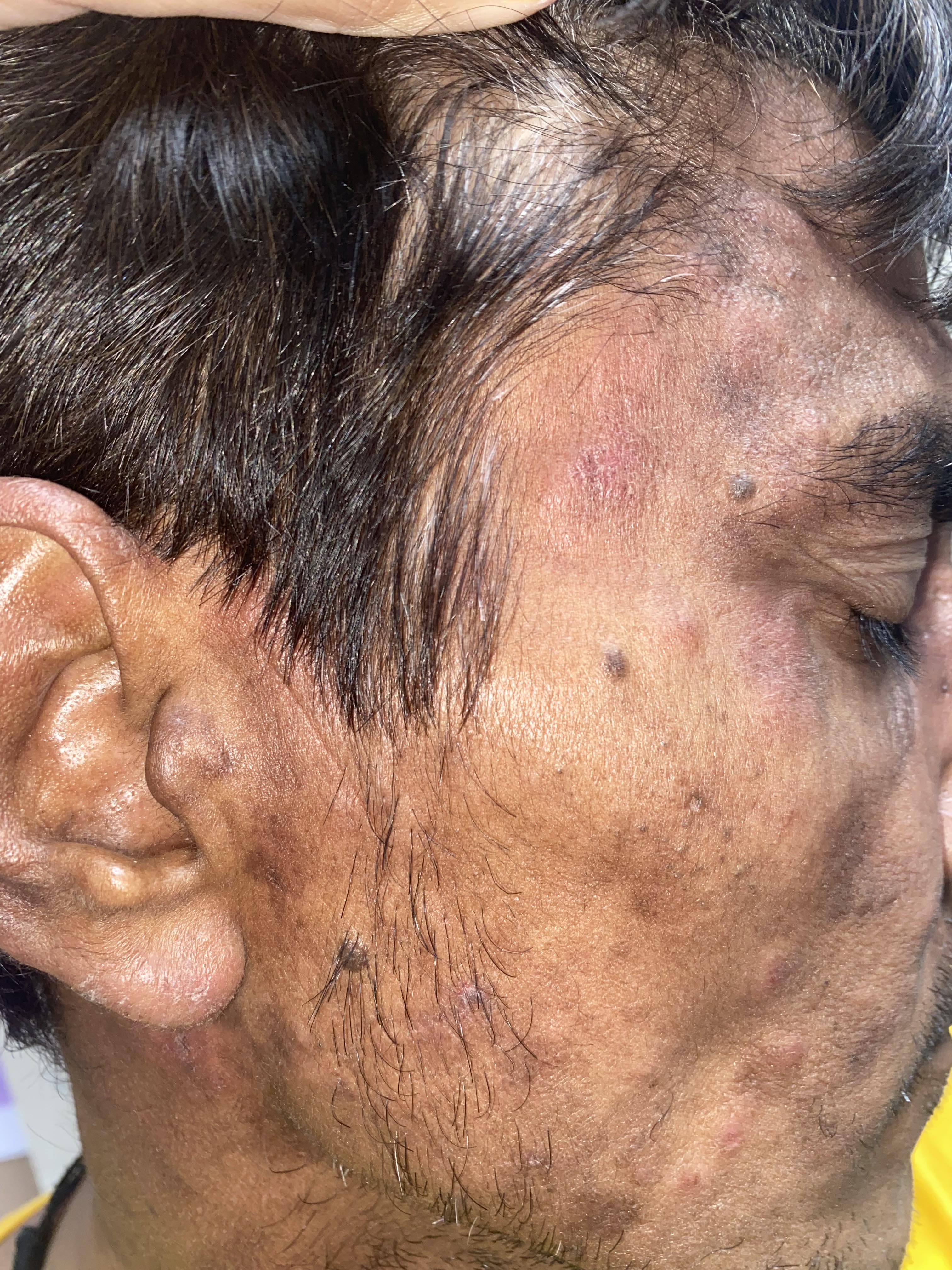
Identify the disease from the four pictorial clues. There is extra credit for an explanation of the fourth clue.
Click here for the answer
March 22, 2023
Suman Patra
 Click on the image to enlarge
Fill in the crossword puzzle with commonly used acronyms from dermatopathology and a little beyond.
Clues:
ACROSS
1: A bothered interface with deeper infiltrate
4: A special stain occasionally needed in skin
5: The one who recommends
DOWN
1: Thin blisters over the folds: it's IgA in IF but what is the target?
2: Necrotizing granulomas in skin are really few and far between
3: Hold your cuff tight both up and down; round
Click here for the answer
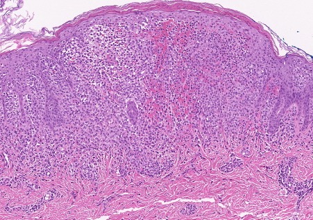
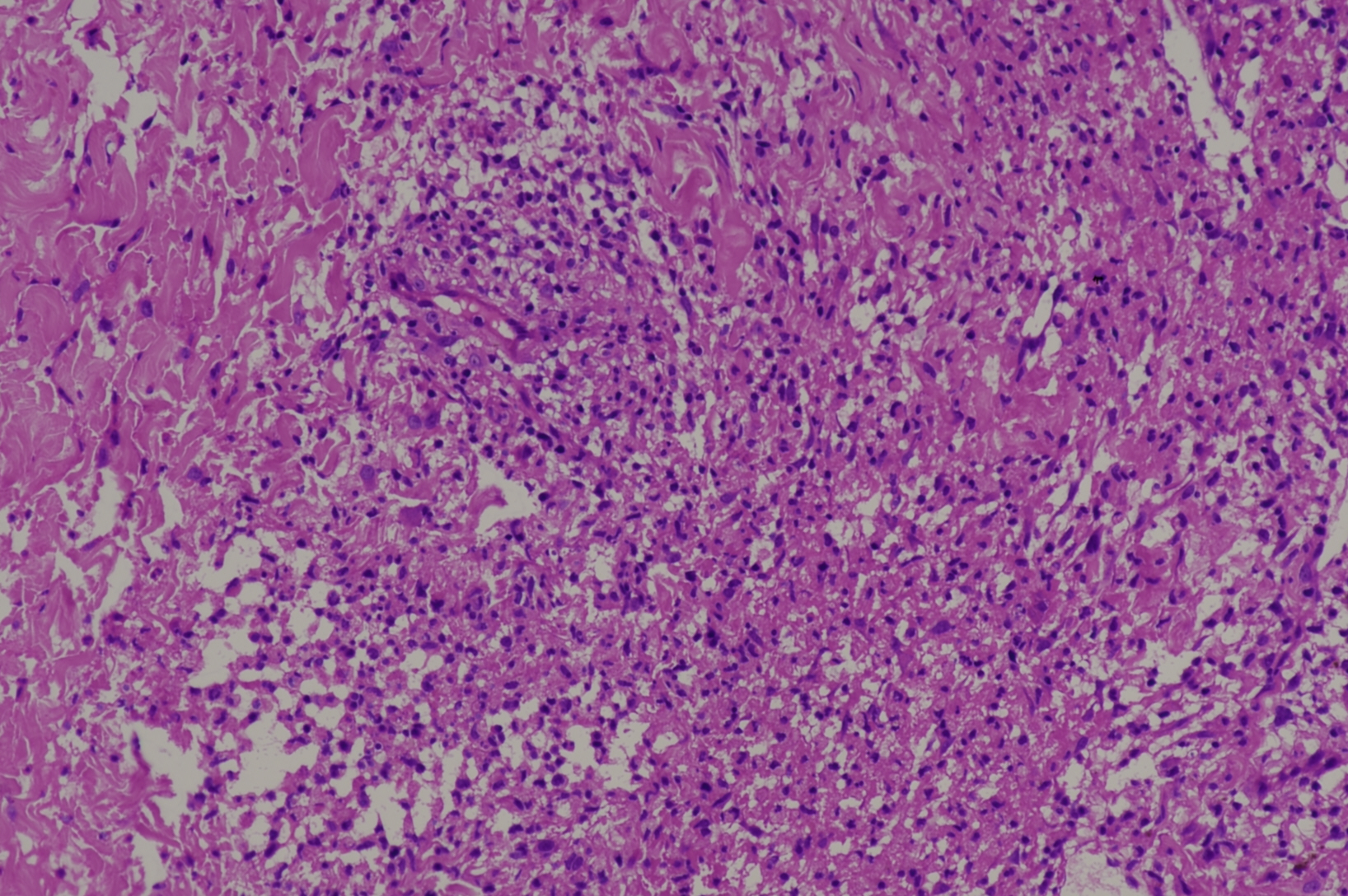
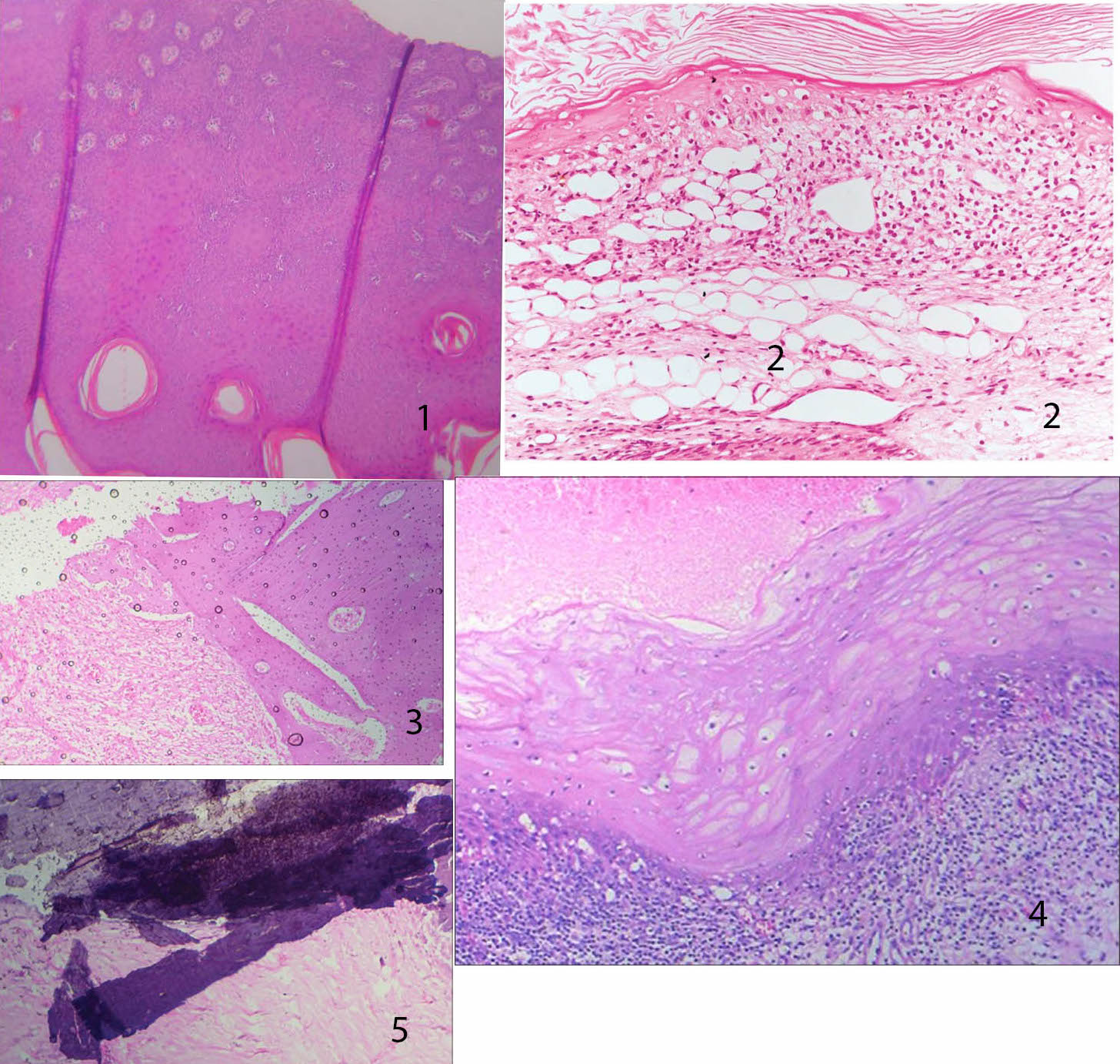
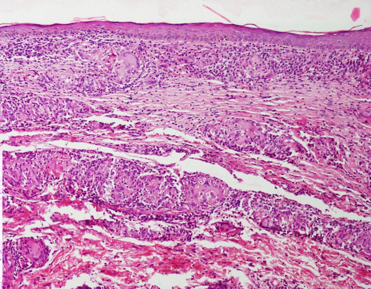
February 23, 2023
Suman Patra
 Click on the image to enlarge
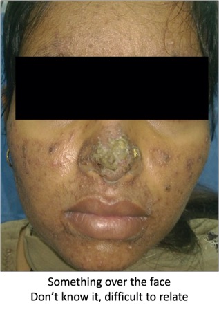
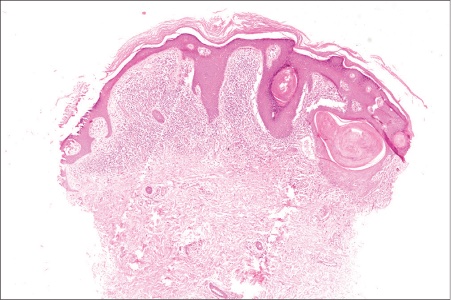
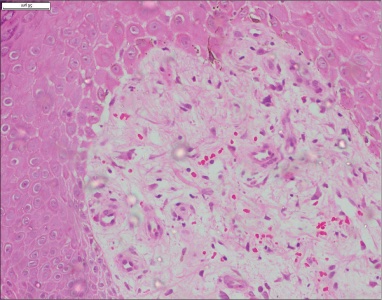
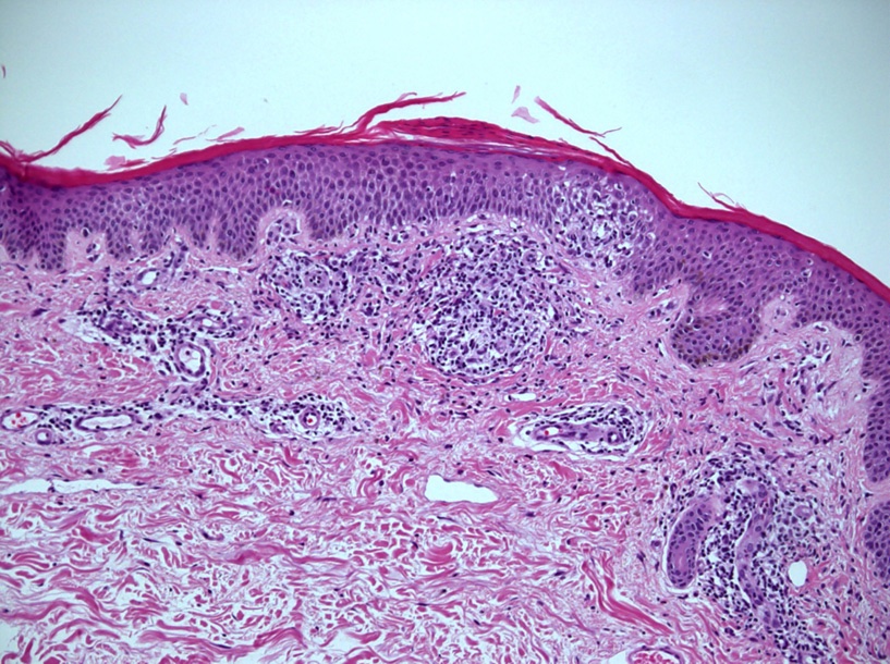
Identify the disease from its histopathological features in the photomicrographs. There are other pictorial clues in the collage.
What is your diagnosis?
Click here for the answer
January 22, 2023
Suman Patra
 Click on the image to enlarge
Identify the common clue or the disease related to the pictures above.
Click here for the answer
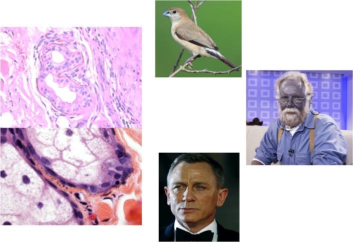
December 23, 2022
Suman Patra
I’m darker than the rest
Little distant from the top
Kind of ‘mixed’ of the lot
Bit ‘fatty’ over the ankle.
My distant cousin, though boring
Always has a better 'stori' to tell
Who am I?
Click here for the answer
November 17, 2022
Suman Patra
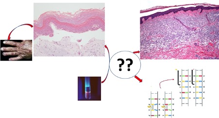 Click on the image to enlarge
The picture shows various processes or findings. The circle at the center of the picture is the missing link between all of them. What is it? What are the individual findings that lead to the answer?
Click here for the answer
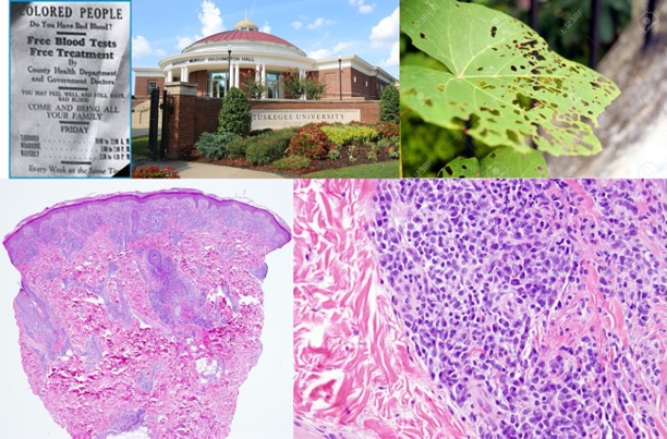
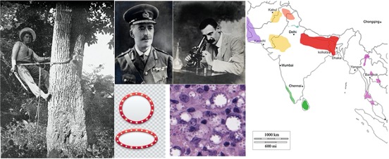
October 20, 2022
Suman Patra
Identify the disease in question from clues in the pictures
 Click on the image to enlarge
The ones who suffered in the past (pic 1)
The great minds who figured it out (pic 2a, 2b)
A stretch of imagination to find a mimic (pic 3a, 3b)
Places still we get quite a bit of it (pic 4)
Click here for the answer
September 20, 2022
Suman Patra
You have to identify the disease from the clues. Some clues are hidden in the text below. Second clue is in the photomicrograph.
Clue 1
Look innocuous over the skin
Don’t ignore me! Not an infant anymore…
Geography can give you the clue
Come to the ‘court’, ‘ace’ it through
Clue 2
 Click on the image to enlarge
Click here for the answer
August 22, 2022
Suman Patra
Identify the disease with four pictorial clues.
(Picture courtesy: Dr. Neetu Bhari)
Click on the images to enlarge
Click here for the answer
July 18, 2022
Suman Patra
Find out the common link between the five pictures provided:

Click on the images to enlarge
Click here for the answer
June 16, 2022
Suman Patra
We have three clues to identify the skin lesions here.
The first clue is in the text below:
I burn under the sun, joint that bites you
SLICC I fulfil, on the skin you got these
Polymorphs in plenty, interface might get
Looking for bullae, ‘ll never get me correct.
The second and third clues are in the clinical and histopathology pictures:
 Click on the images to enlarge
Click here for the answer
May 16, 2022
Suman Patra
Recognize me from the clues hidden in these lines:
The last thing Tyrannosaurus saw
Coming from the sky.
Smaller scale, we see it though
Not very own to the one thou try
Foothills are where we see it best
Moves in line, in sphagnum it rests.
Click here for the answer
April 27, 2022
Suman Patra
 Click on the image to enlarge
1. Identify the skin disease in question correlating all the four pictures.
2. Explain how they are related to the disease.
Click here for the answer
March 22, 2022
Suman Patra
Look carefully at the photomicrographs below.
 Click on the image to enlarge
1 What do you think is the common link between these seemingly unrelated photomicrographs?
2 How are image 2 and image 4 different from the rest?
Click here for the answer
February 26, 2022
Suman Patra
Identify the disease with the help of two clues: text and image.
Clue 1
Beautiful are they, in whom I stay,
Far from you, by the Pacific it may;
Vessel it is, sometime I creep
Symptoms are less than what it seems
You have to 'fight' to pick me right.
Clue 2
 Click on the image to enlarge
Click here for the answer.
January 26, 2022
Debajyoti Chatterjee
An 8-year boy presented with diffuse swelling of both the lips with erosions. There was a history of recurrent discharge from ear, lower respiratory tract infections, and multiple soft tissue abscesses from infancy. Genetic analysis revealed a homozygous defect in NCF1 (c.73_74del GT, frameshift, p. Tyr26HisfsX25). A photomicrograph of the lip biopsy appears below:
 Click on the image to enlarge
A What is the likely diagnosis?
B What other laboratory test(s) can be done to confirm the diagnosis?
Click here for the answer
December 24, 2021
Debajyoti Chatterjee
 Click on the image to enlarge
What instrument is shown in the picture above?
What is the principle of its use?
Click here for the answer
November 25, 2021
Debajyoti Chatterjee
 Click on the image to enlarge
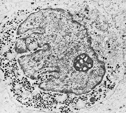
Electron microscopy of an epidermal cell is shown above.
Describe the findings and recognize the cell.
What immunohistochemistry can be used to identify this cell?
Click here for the answer
October 23, 2021
Debajyoti Chatterjee
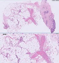
A 63-year female presented with acute onset abdominal pain and nodular necrotic lesion in left forearm. On systemic investigation, she had anaemia. A skin biopsy from the left forearm was performed. A low and high-power image of the biopsy appear below.
a What special stain(s) should be done to establish the nature of the material?
b What do you expect on direct immunofluorescence?
 Click on the image to enlarge
Click here for the answer
September 22, 2021
Debajyoti Chatterjee
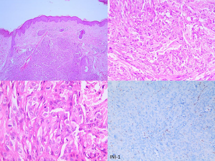
A 33-year male presented with a large nodular lesion in the anterior abdominal wall. A skin biopsy was performed, a representative photograph has been provided. The tumor cells were positive for cytokeratin, vimentin, calponin, S-100, p63 and negative for HMB-45, CD31, CD34, CD68 and CD45.
a. Interpret the INI-1 immunostain as shown in the figure.
b. What is the most likely diagnosis or the closest differential diagnosis?
 Click on the image to enlarge
Click here for the answer.
August 23, 2021
Debajyoti Chatterjee
A 53-year male presented with multiple cutaneous nodules on ear, face and back. On systemic examination, he was found to be hypertensive. A skin biopsy was performed.
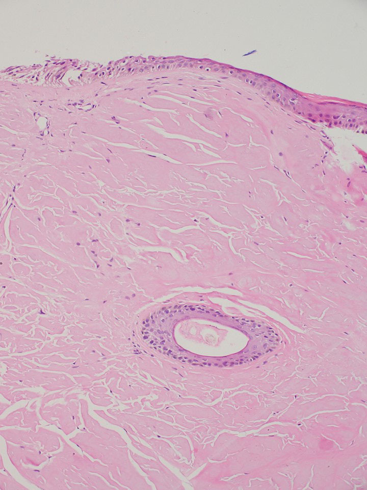 Click on the image to enlarge
a.What histochemical stain will you perform to confirm the diagnosis?
b.What special instruction will you provide to your technician?
Click here for the answer
July 19, 2021
Debajyoti Chatterjee
What is the significance of this image in dermatopathology:

Click here for the answer
June 21, 2021
Debajyoti Chatterjee
A 13-year boy presented with a reddish nodule measuring 1.5x1 cm on left cheek. On examination, he also had left preauricular lymphadenopathy.
The nodule was biopsied::
 Click on the image to enlarge
The tumor cells are positive for CD45, CD30 and ALK-1, and negative for CD3, CD20, CD2, CD5, CD7 and PAX5.
What is your diagnosis? How will you further investigate the patient?
Click here for the answer
May 28, 2021
Debajyoti Chatterjee
A 43-year female patient presented with recurrent non-healing ulcers on both lower limbs. She is a school teacher by profession. She also gives the history of a few episodes of haematuria. She was investigated.
Interpret the following test and suggest further steps for confirmation.
 Click to enlarge
Click here for the answer
April 21, 2021
Debajyoti Chatterjee
A 56 year female patient presented with diffuse mucocutaneous erosion with ulceration. Examination of skin biopsy showed a suprabasal bulla with few acantholytic cells and dense lymphoid infiltrate in the upper dermis. On CT scan abdomen, a large pelvic mass was identified. A diagnosis of paraneoplastic pemphigus is suspected. What is the confirmatory investigation and what is the suitable sample to be taken from the patient for that investigation?
Click here for the answer
March 15, 2021
Debajyoti Chatterjee
In the image below, identify the structures and their relationship in dermatopathology:
 Click to enlarge
Click here for the answer
February 15, 2021
Debajyoti Chatterjee
Identify me:
When in white light, I’m not seen
Placed in blue, I turn green
I’m found attached to antibody or protein
Hit at 495, I jump to 520 as a routine
Click here for the answer.
January 15, 2021
Debajyoti Chatterjee
Which of the following statements about Markel cell carcinoma (MCC) is false?
a. MCC is always CK20 positive
b. MCC can arise within other adnexal tumors
c. It is an indolent tumor, and behaves less aggressively compared to melanoma.
d. MCC can show melanocytic differentiation
e. Some cases of MCC can show basal cell carcinoma (BCC)-like differentiation with peripheral cleft formation
Click here for the answer.
December 15, 2020
Debajyoti Chatterjee
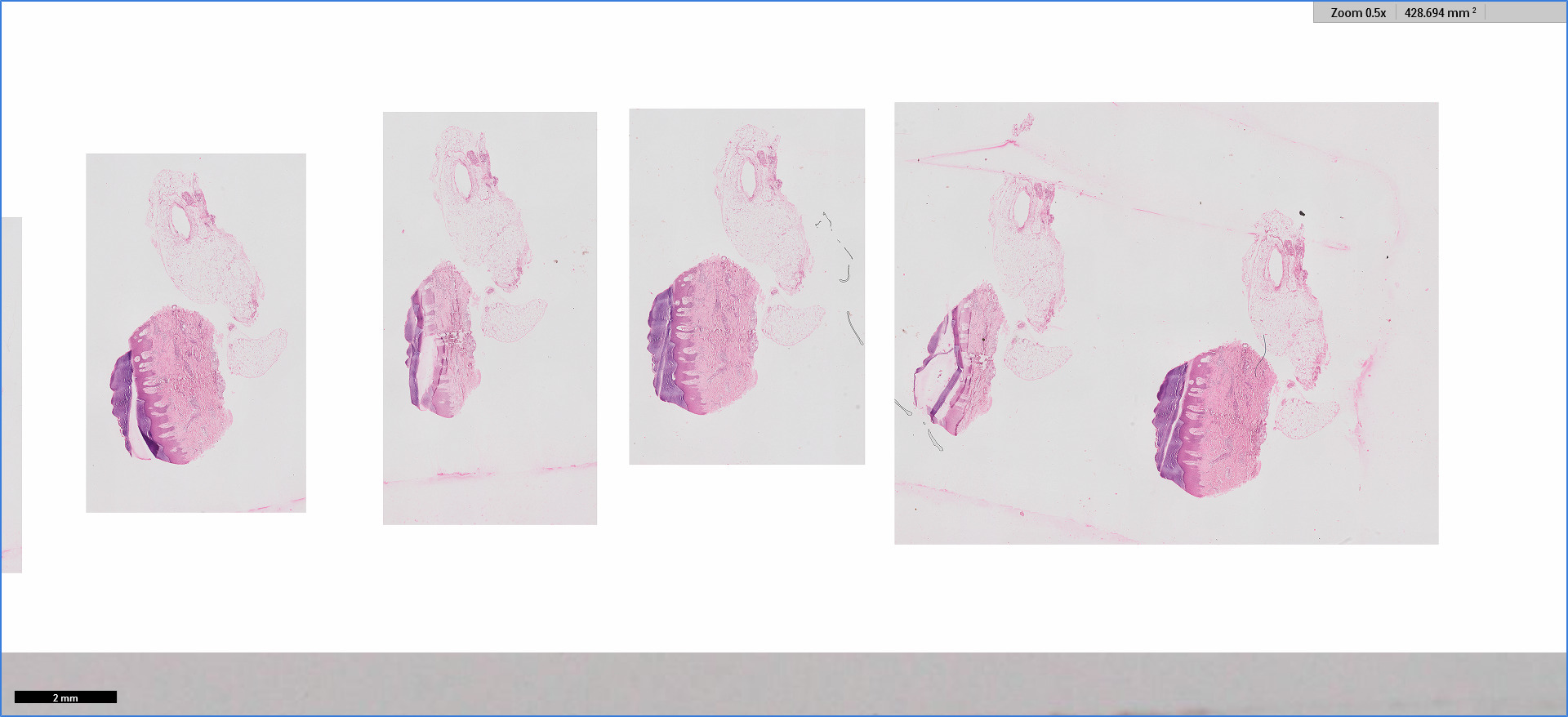 Click to enlarge
This is the photograph of a scanned slide showing serial sections of a skin biopsy. The slide contains some artifact(s). Identify the artifact and its cause.
Click here for the answer.
November 15, 2020
Debajyoti Chatterjee
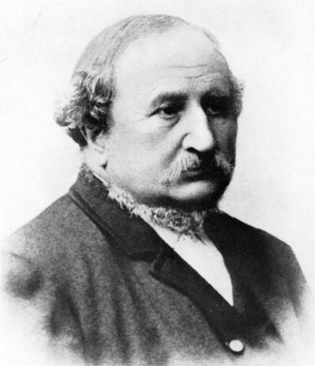 Click to enlarge
Identify this person and name one of his contributions.
Click here for the answer.
October 15, 2020
Debajyoti Chatterjee
A 9-month child presented with recurrent blisters since birth and friable skin. A biopsy is taken.
 Click to enlarge
1 What stain has been used?
2 Interpret the findings.
Click here for the answer.
September 15, 2020
Debajyoti Chatterjee
A 45-year-old female presents to her dermatologist with a 2 cm papule on her back. She had no other comorbidities. A biopsy is performed which shows sheets of atypical large cells in the dermis. These cells are epithelioid in shape, had abundant pale eosinophilic cytoplasm and exhibited frequent mitosis (5-6/ 10 HPF). These cells were immunopositive for S100, CD1a and focally for CD68, while being negative for Langerin, CD34, HMB45, keratin, CD3, CD20, CD30, CD99 and MelanA.
What is the most likely diagnosis?
What do you expect on electron microscopy examination?
Click here for the answer.
August 15, 2020
Debajyoti Chatterjee
A 40 year female presented with multiple non-healing ulcers on both forearms and difficulty in eating for three months duration. On oral examination, she had oropharyngeal ulcers. She gave history of renal transplantation for IgA nephropathy 3 years ago and on immunosuppression since then. There was no lymphadenopathy on clinical examination or imaging. A skin biopsy performed showed dense dermal mixed cellular infiltrate composed of lymphocytes, plasma cells, eosinophils and few histiocytes. Few large binucleated cells were observed which showed positivity for CD30, CD15, but was negative for CD3, CD20 and PAX5.
1 What is the most likely diagnosis?
2 Which investigation will you perform to confirm the diagnosis?
Click here for the answer
July 15, 2020
Debajyoti Chatterjee
A 25-year-old female presents to her dermatologist with a 10 mm pigmented papule on the cheek. A biopsy is performed which shows a wedge-shaped intradermal lesion composed of large, pigmented epithelioid, spindled, and dendritic melanocytes with abundant heavily pigmented melanophages. Mitosis is infrequent. What genetic alteration is most commonly associated with this lesion?
Click here for the answer
June 15, 2020
Debajyoti Chatterjee
A 56-year male presented with multiple firm to hard cutaneous nodules. The dermatologist suspected cutaneous metastasis. On radiological evaluation, a mass is detected in the hilum of left lung. Histological evaluation of the cutaneous nodule shows a poorly differentiated tumor in the dermis, composed of cells arranged in diffuse sheets, with high N:C ratio, coarse chromatin, prominent nucleoli, and frequent mitosis. The tumor shows foci of abrupt squamous differentiation with keratin pearl formation. Tumor cells are negative for p40.
1. What is the most likely diagnosis?
2. Which immunostain will you perform to confirm the diagnosis?
Click here for the answer
May 15, 2020
Debajyoti Chatterjee
An overweight, 63- year male presented with multiple subcutaneous, non-tender, pinkish to reddish nodules for three months. The nodules ranged in size from 5 to 14 mm. On investigations, his uric acid level was found to be elevated (12.5 mg/dl). A biopsy was taken for routine histopathological examination. What do you expect in the histological examination?
a. A lobular panniculitis composed of eosinophils and plasma cells.
b. Needle shaped crystals in the subcutaneous fat surrounded by macrophages and giant cells
c. Needle shaped crystals in the subcutaneous fat surrounded by neutrophils
d. Amorphous pale eosinophilic fibrillary material in the subcutaneous fat, surrounded by lymphocytes and macrophages
Click here for the answer
April 15, 2020
Debajyoti Chatterjee
A 29-year woman presented with abdominal pain. On evaluation, there were multiple discrete cutaneous nodules in both upper extremities. She underwent exploratory laparotomy and removal of the abdominal tumor. The gross and microscopic appearance of the tumour appears below.
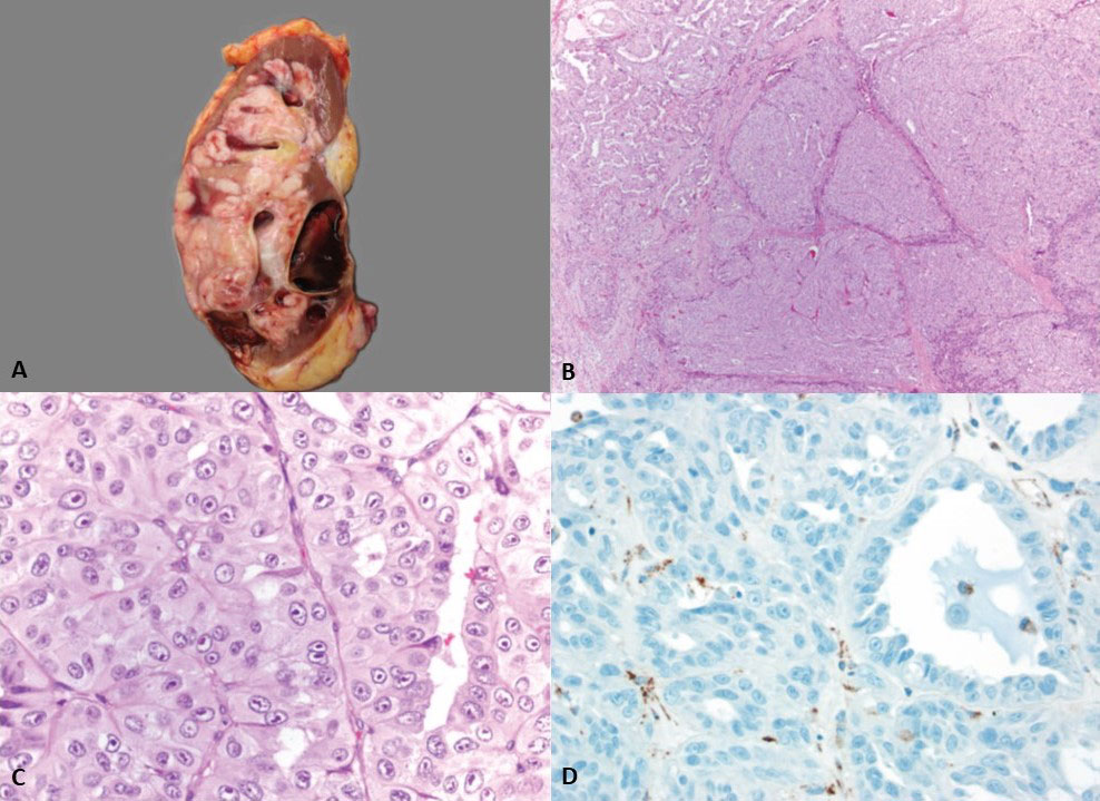 Click on the thumbnail to see a larger image
1 What do you expect on histological examination of the cutaneous nodule?
2 Which immunostain has been shown in figure D? It shows loss of expression in the tumor cells (endothelial cells being positive serve as internal control).
Click here for the answer.
March 15, 2020
Geethanjali Gude

In the picture above, there are two images. Try to relate these two images, which gives clue towards a particular structure/ cell/ event related to dermatopathology. Identify the structure/ cell/ event.
Click here for the answer.
February 15, 2020
Debajyoti Chatterjee

In the picture above, there are two images that suggest a particular structure/ cell/event related to dermatopathology
1. Identify the structure/cell/event
2. How is this structure/cell/event is related to dermatopathology?
Click here for the answer.
January 15, 2020
Debajyoti Chatterjee

In the picture above, there are two images that suggest a particular structure/ cell/event related to dermatopathology
1. Identify the structure/cell/event
2. How is this structure/cell/event is related to dermatopathology?
Click here for the answer.
December 15, 2019
Divyalakshmi C
Identify the disorder described in the riddle:
Born from the wit of an American
My ephemerality is known
Heat, sweat, UV-light
Invite me right
Itchy rash on the chest
Save this old man with your best
Breaking the cement is my habit
Look at the pattern to crack it
Histological faces are many
My pink goggled friends often accompany
Catch me if you can!
Click here for the answer.
November 15, 2019
Divyalakshmi C
Identify the disorder described in the riddle:
Unna’s pattern on the peak
The dermis with a fiery red leak
The lid is open but no tea in pot
Dyskeratosis seen on the major dot
The tilted mounds is the catch
Makes it unique from clinical match
What are we talking about?
Click here for the answer.
October 25, 2019
Divyalakshmi C
Identify the disorder described in the riddle:
I am progressive but benign
in the color of wine
Grouped red- purple beads on this woman's hands and feet
Immunohistochemistry takes the beat
I have proliferating vessels in mid-dermis
But I am not a vascular tumor
I have collagen bundles thick
But I am not sclerotic
Scattered angulated cells with peripheral nuclei in mid-dermis
But I am not granulomatous
Vimentin, factor XIII positive bizzare cells
But CD 31, 34, S-100 negative cells
Who am I?
Click here for the answer.
September 15, 2019
Divyalakshmi C
Identify the disorder described in the riddle:
Infections, immunodeficiency or autoimmunity
I follow these without any rationality
Nodes are my lovely host
But the skin also bears the cost
I Dam up the milky channel
Because that’s where I love to dwell
Lumps and bumps is all you have to face
But many a times I leave without a trace
Give me one, I say “No”
Give me 100, I say “S”
Dermal infiltrate of cells with nuclei that is vesicular
A keen watch can fetch you cells like the ‘intact pearls within oyster’
Who am I?
Click here for the answer.
August 15, 2019
Divyalakshmi C
Identify the disorder described in the riddle:
I take away the smiles
Bowel full of turmoils
The horseshoe is my wish
I give it to you, that blush
In the beginning, its all clear
Later I grow down with a pallor
You lose all your charm
Give me what I need, and I am calm!
Who am I?
Click here for the answer.
July 15, 2019
Divyalakshmi C
Identify the disorder described in the riddle:
An asymptomatic eruption of adulthood
Its description made the Germans proud,
Keratotic papules with scales that prick
May bleed when given a kick,
Dermal infiltrates tend to have a lichenoid pattern
Sometimes the Sezary cell-like appearance helps you discern,
Mind the thick pink roof
Although compressed bricks, a better proof,
Lack of cement on a closer view
And you have the perfect cue!
Click here for the answer.
June 15, 2019
Rita Bhatia
Identify the disorder described in the riddle:
Nothing short of an expert’s handicraft,
The diverse morphology is a class apart.
Histological findings are also a novelty,
The only constant is ‘accessibility’.
Epidermal necrosis resembles mummification,
Multinucleated keratinocytes lack chromatin margination,
With so much ongoing and little inflammation,
Trick appears to be getting high on information.
Haemorrhage, fat necrosis and ruptured collagen may drive you cynical
In this disease, ‘atypical is typical.’
Click here for the answer.
May 15, 2019
Divyalakshmi C
Identify the disorder in the riddle:
Scavenger system defect
Skin, hair, eyes have its effect
I resemble an albino
Pulmonary fibrosis and infection can kill me you know!
Granulomatous plaques on vulva and peristomal skin
Call me granulomatous disease, it is a sin
My colon when cut
Resembles treated Leper’s gut
Who am I?
Click here for the answer.
April 15, 2019
Divyalakshmi C
Identify the disorder in the riddle below.
I live in a den, sitting on side wall
You may hesitate to take a call
White opalescent hue
When in doubt, margins are a clue
4 and 13 are my favourite numbers
I do affect other family members
Thick velvety coat I wear
When stained looks clear
I do no harm
Pink clumps in close view are my charm!
Click here for the answer.
March 15, 2019
Divya Chandrasekharan
Identify the disorder in the riddle below.
I was born in the land of the rising sun
Aberrant notch that fires the gun
Hyperpigmented macules that darken with time
I can even escape a crime
Pigment cells in the basal layer
Yet no spill creates a fear
Click here for the answer.
February 15, 2019
Divya Chandrasekharan
Identify the disorder in the riddle below.
The mouse’s favourite meal
Fed by the master’s zeal
The lesion has a rubbery feel
Pain becomes a big deal
Look deep in the section
Lest you miss this condition
Normal architecture of fat cells fall
With chronic inflammatory cells, lipid laden macrophages in the wall!
Click here for the answer.
January 15, 2019
Divya Chandrasekharan
Identify the disorder in the riddle below.
Giant ringworms that makes you fear,
Solar rays get in gear
Mimics tuberculoid leprosy
Necrobiosis and granuloma is all that you see
As it erupts on exposed sites,
Pruritus and irritation is what patient fights
Usually seen in blacks,
Note that mucin and central zone elastic tissue it lacks!
Click here for the answer.
December 15, 2018
Riti Bhatia
Spot the odd one out among the following disorders on the basis of histopathological findings:
ILVEN, actinic keratoses, linear EHK, and nevus sebaceous
Click here for the answer.
November 15, 2018
Riti Bhatia
Identify the disorder in the riddle below.
First described by a Hungarian dermatologist,
The names of this disease are too many to enlist.
The dilemma is not just historical,
But also clinical and histopathological.
Reticulate plaques and seborrheic dermatitis,
Histopathologically mimics classical interface dermatitis.
Renal disease and lymphoid neoplasm may develop in a few,
Look for deeper infiltrates and plasma cells for a clue.
Click here for the answer.
October 15, 2018
Riti Bhatia
Identify the disorder in the riddle:
Itchy benign intertriginous eruption,
Equally fascinating is the reaction pattern.
Look for clues in the dead layer,
You may find something which should not be there.
Sweat, diaper or some unknown effect,
Many share an atopic gene defect.
Click here for the answer.
September 15, 2018
Riti Bhatia
Identify the histopathological pattern in the riddle:
Chronic itchy papules or circinate plaque,
In this histopathological subset, age is no bar.
The disorder in Japanese is not new,
When in immunosuppressed, this is a clue.
Dense infiltrate of lymphocytes, eosinophils and mast cell,
When in sebaceous glands, does it ring a bell?
Click here for the answer.
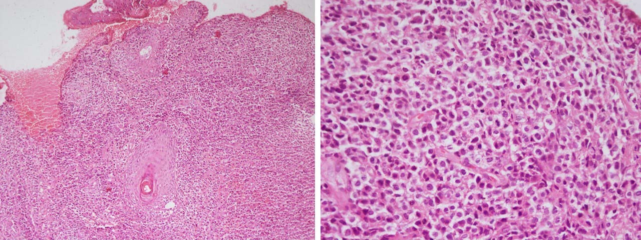
August 15, 2018
Riti Bhatia
Single pink nodule often on the face,
That can grow with a rapid pace.
Time of stagnation will frequently come,
'Self-healing malignancy' is just a pun.
Proliferating large cells with pinkish hue,
Infiltration with 'pink' cells gives a cue.
It can proliferate in and out,
Look at the centre to shed your doubt.
One may ‘overcall’ or ‘undercall’,
Clue lies in the way this tumor will evolve.
Spot the diagnosis in the riddle.
Click here for the answer.
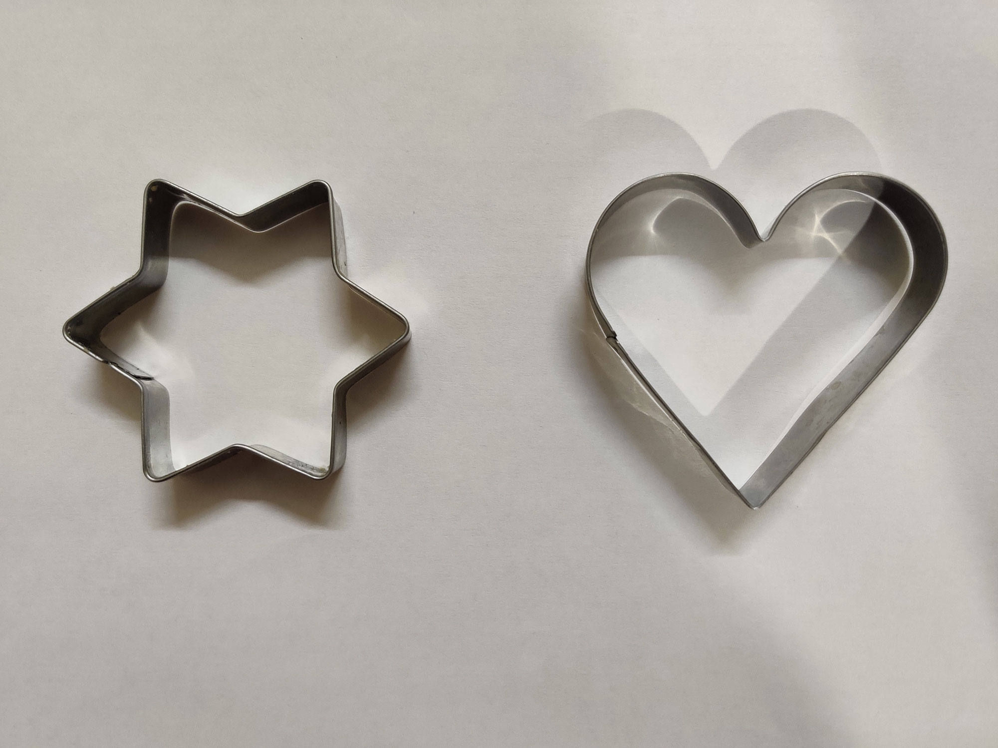
July 15, 2018
Meenakshi Batrani
Spot the disorder in the riddle:
I was born bright red, but I have grown up to have dry horn and torn hair
I always carry golf-tee and a ball
I keep sneezing in the hall
Guess the name if you can recall
Click here for the answer.
June 15, 2018
Riti Bhatia
Papules, pustules and crateriform exudative plaques
With neutrophilic microabscesses and epidermal hyperplasia as hallmarks
Medications, environment and diet are all to blame
Sherlockian history should be the aim
Tumor and infection may be what you think
Pause and ask for what they drink
Although many presidents’ favourite
Stop this drink and see the lesions abate
What's your diagnosis?
Click here for the answer.
May 15, 2018
Vishal Gupta
Spot the odd one out:
Granuloma faciale
Rosai-Dorfman disease
Kimura’s disease
Erythema elevatum diutinum
Click here for the answer.
April 15, 2018
Riti Bhatia
One of those wonderful misnomers,
With clinical and histological bummers.
See early and get lured
For these neutrophils, dust and fibrin are obscure.
Wait for changes to develop
With dense eosinophils and neutrophils, fibrosis will envelop.
This solitary plaque will keep you immersed
Hideously neglecting angiocentric laryngeal whorls.
Click here for the answer.
March 15, 2018
Riti Bhatia
Itchy velvety plaques which are truncal,
in immunosuppressed patient will make you stumble.
Look in the epidermis for abnormal cell
Does peacock plumage ring a bell?
Trichospinulosa and Merkel carcinoma are connecting link,
This eruption will surely make you think.
Click here for the answer.
February 15, 2018
Riti Bhatia
Spot the odd one out, with reasons:
Basal cell carcinoma
Extra-mammary Paget’s diseas
Trichoepithelioma
Merkel cell carcinoma
Fibroepithelioma of Pinkus
Click here for the answer.
January 15, 2018
Geeti Khullar
I come on forehead as waxy and white,
Plugged crystals behold your sight.
Yeast or bacteria may be found,
Though neutrophils also surround.
Incidentally present near a tumor,
Alcian blue will make you wonder.
Click here for the answer.
December 15, 2017
Riti Bhatia
Spot the odd one out among the following on the basis of histopathological findings and give your reason:
Lichen striatus
Prurigo pigmentosa
Syphilis
Cutaneous lymphoid hyperplasia
Insect bite hypersensitivity
Necrobiosis lipoidica
Click here for the answer.
November 15, 2017
Riti Bhatia
Ashy clues and greenish hues,
Will give astute eyes, ‘the blues’
Tiny dots in eccrine glands, vessels and membranes
these ‘stars in heaven’ will jog your brains.
Identify the phenomenon or pattern being referred to.
Click here for the answer.
October 15, 2017
Riti Bhatia
Single, many or only on a hair follicle,
This horny stack is not just one clonal disorder.
Look for the clues beneath,
Step cuts may be all you need.
Seen in a number of disorders,
Inflammatory, autoimmune, benign and malignant.
Gentian violet can help unveil the borders,
For biopsy site is what matters in the prototype clonal mutant
Identify the phenomenon or pattern being referred to.
Click here for the answer.
.
September 15, 2017
Riti Bhatia
Look through the lens closely,
lurking in subcutis are these jagged clefts,
where the neutrophils came early,
an enzyme is what they left.
This cluster has many cells,
What you are looking for is something else.
Hallmark of a reactive dermatoses,
Are they forgotten in the glory of giant cells?
Identify the histopathological structure described.
Click here for the answer.
August 15, 2017
Riti Bhatia
Sickle bodies, scars and beading,
you will look for them all.
This leonine facies has many differentials,
But the voice says it all.
When too early for onion skinning
Look at glands and vessels for a clue
PAS, colloidal iron and alcian blue
May bring your ‘pale’ intuition true!
Discovered by two Viennese,
What is the name of this disease?
Click here for the answer.
July 15, 2017
Riti Bhatia
Dilated cystic spaces in sclerotic stroma,
Leave you mesmerized with a ‘comma’
Provocative shapes of epithelial islands
Involucrin, CEA, BerEP4 and CK20 are all their stains.
Perineural invasion, horn cysts or retraction cleft
Are clues to some of them in this ‘patterned set’
Click here for the answer.
June 15, 2017
Riti Bhatia
Large cells with ample pale cytoplasm,
small white patches on skin,
Will you think of a neoplasm,
and stain it with PAS, EMA and cytokeratin?
When in doubt,
look for ‘the line’
Biopsy may give you second thoughts
what is your take, malignant or benign?
Click here for the answer.
May 15, 2017
Riti Bhatia
Light or dark on skin, pale in biopsy always.
Look for me superficially, horizontal are my ways.
I may be alone or in a syndrome, upper body is where I am seen.
When stuck with peripheral palisading, you will find the answer with orcein.
What is your diagnosis?
Click here for the answer.
April 15, 2017
Riti Bhatia
What is the connecting link between progressive symmetric erythrokeratoderma, papillomatosis cutis carcinoides and acrogeria?
Click here for the answer.
March 15, 2017
Geeti Khullar
Which of the following is the odd one out and why?
1. Post kala-azar dermal leishmaniasis
2. Erythema elevatum diutinum
3. Mycosis fungoides
4. Dermatofibroma
Click here for the answer.
February 15, 2017
Meenakshi Batrani
Identify the common morphological (gross or microscopic) term for the following entities:
1. Elastofibroma
2. Trichorrhexis nodosa
3. Actinomycetoma
4. Lipoid proteinosis
5. Atypical mycobacteria
Click here for the answer.
January 15, 2017
Meenakshi Batrani
Identify the common morophological term for the following entities:
1. Borderline leprosy
2. Erythema elevatum diutinum
3. Lipoid proteinosis
4. Perineuroma
5. Myopericytoma
Click here for the answer.
December 15, 2016
Riti Bhatia
From nevi to lentigenes to melanoma,
I resemble all those big names.
Young people are my favourite,
I go away without giving any pains.
The upper you go,
Better will be the clues.
Stain me with Perls or Benzidine
Which stain will you choose?
Identify the condition and stain used.
Click here for the answer.
November 15, 2016
Riti Bhatia
What is the common term that links these conditions: hidroacanthoma simplex, pachyonychia congenita, blue nevus and a rare form of ectodermal dysplasia with keratin mutation?
Click here for the answer.
October 15, 2016
Vishal Gupta
Based on histopathologic features, which one among the following is the odd one out and why?
1. Erythema toxicum neonatorum
2. Transient neonatal pustular melanosis
3. Acropustulosis of infancy
4. Miliaria pustulosa
5. Neonatal acne
Click here for the answer.
September 15, 2016
Riti Bhatia
What is the connecting link between multiple basal cell carcinomas, Ehlers-Danlos disease and focal dermal hypoplasia?
Click here for the answer.
August 15, 2016
Riti Bhatia
Which of the following is the odd one out and why?
CHILD syndrome
Verruciform xanthoma
Hidradenitis suppurativa
Fox-Fordyce disease
Click here for the answer.
July 15, 2016
Rajalakshmi T
Which is the odd one out and why?
1. Cylindroma
2. Spiradenoma
3. Fibrofolliculoma
4. Trichoepithelioma
Click here for the answer.
June 15, 2016
Khalid Al Aboud
Which is the wrong number among these and why?
CD 117, CD 2, CD 14, CD 25
Click here for the answer.
May 15, 2016
Khalid Al Aboud
I demonstrate dusky dappled disfigurements
and dark dot depressions,
disclosing digitate downgrowths delving dermally.
Who am I?
Click here for the answer.
April 15, 2016
Contributed by Rajalakshmi T
I favour the head and neck,
I show continuity with infundibula,
I am populated by dark and pale cells,
I show ducts, but only within the pale areas,
Necrosis is my good friend.
Who am I?
Click here for the answer.
March 15, 2016
Contributed by Meenakshi
Batrani
I have a red face with a goggle!
Drugs make me sparkle!!
Burning in the flame, I always take the blame!!
Insects give me fame!
Just guess my name!!
Click here for the answer.
February 15, 2016
Contributed by Meenakshi
Batrani
I am the freshener of the mouth, brightening many a cloud!
Excess of me you take, will make you like a slate!!
Who am I and what condition do I cause?
Click here for the answer.
January 15, 2016
Contributed by Geeti Khullar, Saurabh Singh
Although my founder literally adorns the ocular cilium; in life horny and not so pretty I am.
I may appear to love solitude, multitude, genetics or may even be reactive; I tend to embellish the senile more than the active.
Closer you look and the deeper you tread; I get ducts embedded in strings-strands, and vessels and mast cells in the bed.
Behold my often benign innocent ways, I may dawdle to cancery lanes.
Don’t put thy intellect to shame, what shall be my lengthy name?
Click here for the answer.
December 15, 2015
Contributed by Rajalakshmi T
What is the descriptive term (microscopic, gross or imaging) common to the following conditions?
1. Melanoma
2. Syphilis
3. Fibromatosis
4. Scurvy
Click here for the answer.
November 15, 2015
Contributed by Rohini Mathias
Which is the eponym common to the three descriptions listed below?
1. Used to characterize and discriminate between various connective and soft tissue elements of a lesion
2. Clinical mimic of angiosarcoma
3. Used to highlight melanocytic lesions
Click here for the answer.
October 15, 2015
Contributed by Rajalakshmi T
All the following statements are true about a particular disease except one:
a. They occur more frequently in blacks
b. They stain positive for Factor XIIIa.
c. They contain dendritic melanocytes
d. They stain variably with CD34.
Which is the condition described above and which statement is incorrect?
Click here for the answer.
September 15, 2015
Contributed by Rohini Mathias
Pick out the histologic metaphor that does not fit, with a justification for your choice:
1. Lump of coal on a pillow
2. Toy soldier appearance
3. Eyeliner sign
4. Bare under belly sign
Click here for the answer.
August 15, 2015
Contributed by Rohini Mathias
Fever and lipodystrophy
growth retardation and hepatomegaly,
Annular lesions with typical facies,
are the clinical features one sees....
there's a mixed dermal and subcutaneous MPO positive infiltrate,
know this bright new syndrome and you're up to date!
What is the disease described above?
Click here for the answer.
July 15, 2015
Contributed by Rohini Mathias
The syndrome characterised by multiple cutaneous neoplasms having a fenestrate pattern of mantle-like cells with clusters of sebocytes at times is also associated with all of the the following, except:
1. Renal oncocytoma
2. Renal medullary carcinoma
3. Chromophobe renal cell carcinoma
4. Papillary renal cell carcinoma
Click here for the answer.
June 15, 2015
Contributed by Rohini Mathias
Pick the odd one out from these metaphorical histologic terms and give the reason for your choice.
1. Shark's tooth
2. Paisley tie
3. Rolls and scrolls
4. Onion skin
Click here for the answer.
May 15, 2015
Contributed by Rohini Mathias
Your patient is a febrile Latino with mulberries in his mouth,
Though the histology looks like cancer, there’s a granuloma without a doubt!
So sail across the oceans and grab hold of the mariners wheel,
Then to us your diagnosis you can confidently reveal….
Which disease is being described here?
Click here for the answer.
April 15, 2015
Contributed by Rohini Mathias
Usually spotted in the Native American and Inuit race,
These lesions are found on a mucosa in the face.
A virus has been incriminated in its etiology,
An axe from the bronze age is what you see on microscopy!
Which condition is this?
Click here for the answer.
March 15, 2015
Contributed by Rohini Mathias
What is the common microscopic term that links the following conditions?
1. Hibernoma
2. Fabry's disease
3. Protothecosis
4. Measles
Click here for the answer.
February 15, 2015
Contributed by Rohini Mathias
Spot the histological odd man out with a suitable reason to support your choice:
1. Fordyce spots
2. Muir - Torre syndrome
3. Pearly penile papules
4. Juxtaclavicular beaded lines
5. Steatocystoma multiplex
Click here for the answer.
January 15, 2015
Contributed by Rohini Mathias
Which condition has all the features mentioned below?
1. Predilection for head and neck region
2. Perineural infiltration
3. Ductal differentiation, with downgrading of duct sizes in the deeper parts
4. Eosinophils in the infiltrate
5. CK-7, CK-15 and CD5 positive cells
Click here for the answer.
December 15, 2014
Contributed by Rohini Mathias
Three tumors and an allergy…what could the link between these be?
1. Spongiotic vesicular dermatitis
2. Rhabdomyosarcoma
3. Squamous cell carcinoma of the cervix
4. Syringoma
With reference to microscopic features, what is the descriptive term common to the above conditions?
Click here for the answer.
November 15, 2014
Contributed by Rohini Mathias
It may be invisible at first says the book,
But those dilated eccrine ducts deserve a second look,
Do take that biopsy at the appropriate time,
Else all will be wasted, and this isn't just a rhyme!
So immerse your hand in water little girl,
Let your diagnosis unfurl!
Which is the disease being talked about here?
Click here for the answer.
October 15, 2014
Contributed by Rohini Mathias
Histologically, who is the odd man out...and why?
1. Sclerema neonatorum
2. Pancreatic panniculitis
3. Post-steroid panniculitis
4. Subcutaneous fat necrosis of newborn
Click here for the answer.
September 15, 2014
Contributed by Rohini Mathias
During immunosuppression I choose to surface...
Lots of plasma cells and histiocytes is what you see....
I take up Perls, Alizarin red and PAS,
That is how you will diagnose me!
a) What is the condition?
b) What are these histiocytes called?
Click here for the answer.
August 15, 2014
Contributed by Rohini Mathias
Here are histological descriptions of 3 different cutaneous entities.
1. Collection of bipolar and dendritic melanocytes in the interstices of dermal collagen, tending to concentrate around appendages
2. Histologic pattern characterized by sharply defined intraepidermal nests of morphologically different cells
3. Focal loss of elastic fibres with normal collagen preceded by an inflammatory lesion
What is the 'name' common to these three?
Click here for the answer.
July 15, 2014
Contributed by Rohini Mathias
Sew up the following clues together to crack this one…what’s your final diagnosis?
1. Gray horses
2. Black with occasional satellites
3. Dendritic and epithelioid cells
4. Predominantly dermal and occasionally deeper extension
5. Adnexotropic
Click here for the answer.
June 15, 2014
Contributed by Rohini Mathias
There's a lot of lipid everywhere you see,
Be it in the keratinocyte or the Jordan's anomaly....
Some are born in hot wax or erythrodermic,
but peripheral nerves, they aren't so thick...
Inherited as an autosomal recessive trait,
A retinoid and dietary restrictions may improve this child's fate!
Identify the disease in the riddle....
Click here for the answer.
May 18, 2014
Contributed by Rohini Mathias
Which among the following features does not fit into the 'Last Week's Sign' ?
1. Parakeratosis overlying basket-weave orthokeratosis
2. Mild epidermal hyperplasia
3. Numerous necrotic keratinocytes
4. Mild dermal inflammation
Click here for the answer.
April 15, 2014
Contributed by Rohini Mathias
Fish out the red herring and give a reason for your choice.
a. Rheumatoid vasculitis
b. Granuloma annulare
c. Wegeners granulomatosis
d. Necrobiotic xanthogranuloma
Click here for the answer.
March 15, 2014
Contributed by Rohini Mathias
People say I’m ‘3’ faced…but I’d best describe myself as ‘dimorphic’ ,
‘Roses’ are my favorite, but better beware of that thorn prick!
I prefer ‘cigars’ to cigarettes and astronomy intrigues me…
Microscopically I can create a cocktail!
Who am I?
Click here for the answer.
February 15, 2014
Contributed by Rohini Mathias
What is the common descriptive term used in the microscopic findings of the following conditions?
a. Ochronosis
b. Pityriasis versicolor
c. Spitz nevus
Click here for the answer.
January 15, 2014
Rajalakshmi T, Inchara YK

Clues:
Across
1. If you want to get rid of this without a hitch, treat the baby and the grandpa for this itch.
4. Foamy, foamy in a sheath, seen in the midst like a wreath
8. Cells around a centre, pink or blue, Gran Ann is your clue
9. Schaumann and Asteroid in their cosy nests, skin deep goes this test
11. Many needles in this haystack, pain in the toes keeps coming back
12. Princess of Prussia, of royal blue blood
Down
2. Cells undergone apoptosis, in this band-like dermatosis
3. What’s your email _ _?
5. Papules and pustules close to hair, spectacled cells everywhere
6. Virchow had a Mexican feast, thrombi in the vessels - how neat!
7. Bees buzzing in Rapunzel’s hair, caused her to exclaim in despair.
10. Rafa grips this like a winner; bumps in the skull, marrow and the live

December 15, 2013
Spot the odd one out:
a) Henoch-Schonlein purpura
b) Atrophie blanche
c) Finkelstein’s disease
d) Granuloma faciale
Click here for the answer.
November 15, 2013
In a skin eruption of short duration, all the following features suggest a drug-related cause except:
a) Basket-woven orthokeratosis
b) Extravasated red cells
c) Wiry collagen in the papillary dermis
d) Many necrotic keratinocytes
e) Dermal oedema
Click here for the answer.
October 15, 2013
Which two tumors are distinguished by their expression patterns of the combination of the immunohistochemical markers Cytokeratin 20, CD10 and Androgen receptors?
a) Trichoblastoma and Trichoepithelioma
b) Proliferating tricholemmal tumor and Squamous cell carcinoma
c) Trichoepithelioma and Basal cell carcinoma
d) Basal cell carcinoma and Squamous cell carcinoma
Click here for the answer.
September 15, 2013
A 45-year-old lady presented with blisters on her limbs. Histopathology showed a cell-poor subepidermal blister with a few dermal neutrophils. Direct immunofluorescence showed deposits of IgG and C3 in a BMZ pattern. Indirect IF on salt-split skin showed deposits localised to the floor of the split.
Which of the following autoantibodies is responsible for these findings and what is the disease?
a) anti BP 230
b) anti BP 180
c) anti laminin gamma 1
d) anti LAD 1
Click here for the answer.
August 15, 2013
I am characterised by dilated infundibula with plugging. Sometimes, I also show cornoid lamellae. But, I am best known for my foamy friends who keep me company around the plugs.
Who am I?
Click here for the answer.
July 15, 2013
I was spotted in the land of the rising sun in 1971. I have a net, but the itching is no fun. Neutrophils flood my epidermis, unlike my roti-hater friend who houses them in the dermis.
Which disease am I?
Click here for the answer.
June 15, 2013
In which condition do you find cystic sebaceous carcinomas?
Click here for the answer.
May 15, 2013
Which disease is characterised by a paranuclear dot pattern of staining with CK20?
Click here for the answer.
April 15, 2013
Which disease shows a chicken-wire pattern along with basement membrane staining on direct immunofluorescence?
Click here for the answer.
March 15, 2013
Which condition is the sandwich sign a clue to?
Click here for the answer.
February 15, 2013
Which disease is characterised by the checkerboard sign?
Click here for the answer.