Answer to Image of the Month May 2013
Submitted by Rajiv Joshi
Incontinentia pigmenti, verrucous stage
In incontinenti pigmenti, the second stage of verrucous papules often begins at sites of resolving vesicles and intermediate stages between the vesicular and verrucous lesions may be seen. The characteristic histologic findings at this stage are numerous dyskeratotic keratinocytes within the hyperplastic epithelium which may be scattered individually or present in whorls. Several eosinophils may still be seen both in the epidermis and the dermis at this stage but as the lesions age the number of eosinophils tends to decrease.
The first image shows numerous dyskeratotic keratinocytes in the mid spinous zone arranged in concentric clumps as well as scattered singly along with exocytosis of few eosinophils in the mildly spongiotic epidermis. The upper dermis is edematous and has several eosinophils. The second image shows numerous eosinophils in the diffuse dermal infiltrate.
The differential diagnoses for this pattern include lichenoid drug eruption, fixed drug eruption and erythema multiforme with unusual presence of eosinophils. The reason/s that the image is not of the above diagnoses is as follows:
1. Lichenoid drug eruption
No parakeratosis is seen. The infiltrate is not strictly lichenoid and has numerous eosinophils. Eosinophils if seen in lichenoid drug eruptions are few in numbers and almost never found in the epidermis along with apoptotic keratinocytes.
2. Fixed drug eruption
While numerous eosinophils and individually necrotic keratinocytes may be seen in this condition, the image shows no melanophages, a finding that is a must in lesions of recurrent fixed drug eruption.
3. Erythema multiforme
Eosinophils as a rule are not seen in erythema multiforme, especially in the numbers seen in these sections. Interface changes of basal cell vacuolization and obscuring of the dermo-epidermal junction which are very common in erythema multiforme are not seen here.
The combination of epidermal hyperplasia with numerous dyskeratotic keratinocytes and numerous eosinophils both in the epidermis and the dermis is very characteristic of the second verrucous stage of incontinentia pigmenti. As the lesion ages, however, the number of eosinophils reduces and may even be absent.
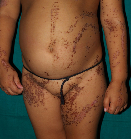 Widespread verrucous papules in a Blaschkoid distribution in a young girl Image courtesy M Ramam