Answer to Image of the Month March 2014
Submitted by Inchara YK, Rajalakshmi T
Granular cell tumor
The images show a dermal-based lesion comprising of intersecting fascicles and sheets of cells with characteristic eosinophilic granular cytoplasm and bland nuclei. The cells are S-100 positive (see image below), affirming their neural character. The first photograph also shows pseudoepitheliomatous hyperplasia of the overlying epidermis, which is a common finding in this tumor and may mimic a squamous cell carcinoma
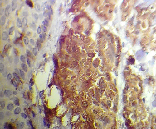 S 100 staining of granular cell tumour