Answer to Image of the Month July 2013
Submitted by Meenakshi Batrani, Asha Kubba
Pityriasis rubra pilaris
The biopsy is from the elbow of a seven year old boy who presented with erythematous scaly plaques on the palms, soles, elbows and knees and erythema on the face and trunk.
Sections show a plugged follicular infundibulum and stratum corneum comprising of alternating orthokeratosis and parakeratosis in both horizontal and vertical directions forming a checkerboard pattern. Epidermis exhibits psoriasiform hyperplasia with broad rete ridges and thick suprapapillary plates and focal hypergranulosis. Mild perivascular lymphocytic inflammation is present in the superficial dermis.
Additional histopathological features which can be seen in some biopsies include mild spongiosis with scattered lymphocytic exocytosis and an occasional plasma cell/eosinophil in the superficial dermal infiltrate.
Pityriasis rubra pilaris requires distinction from psoriasis which it can mimic both clinically and histopathologically. Features favouring psoriasis include confluent parakeratosis, presence of neutrophils forming Munro microabcess and spongiform pustule of Kogoj, hypogranulosis, elongated rete ridges with thinning of suprapapillary plates and tortuous congested vessels in the dermal papillae.
A clinical picture of a similar case appears below.
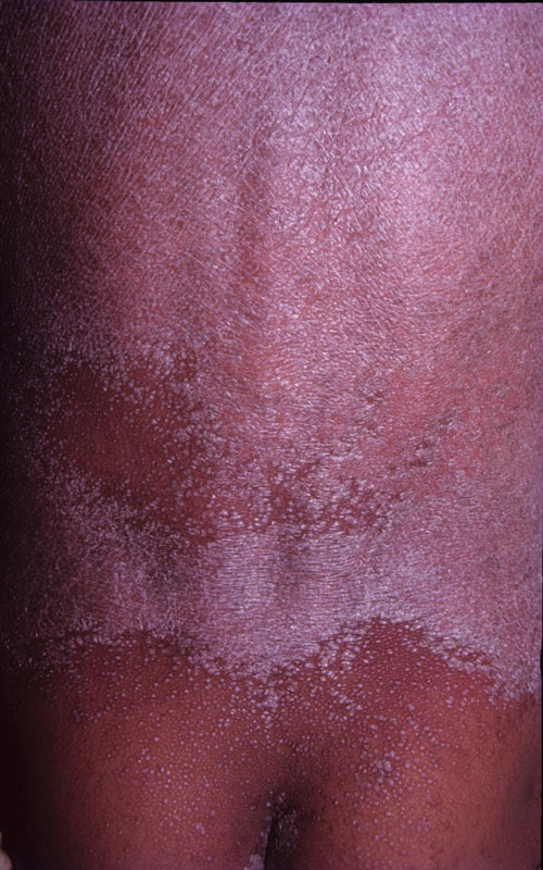
Confluent erythematous scaly plaques enclosing islands of normal skin. Note the tiny follicular papules within the islands and just beyond the margins of the plaques: these are a helpful clinical clue to the diagnosis. (Image courtesy: M Ramam)