Answer to Image of the Month December 2012
Epidermolytic hyperkeratosis
Hyperkeratosis is prominent. There is hypergranulosis. Keratinocytes in the granular and upper spinous layers show vacuolar degeneration with indistinct cell borders and eosinophilic intracytoplasmic inclusions. These changes may extend to varying depths in the epidermis.
Epidermolytic hyperkeratosis can be seen in both epidermolytic ichthyosis ( also known as bullous congenital ichthyosiform erythroderma) and epidermal nevi of the epidermolytic type, disorders which represent the fully expressed and mosaic form respectively of mutations in the genes encoding keratin 1 and keratin 10. Biopsies from the mosaic, nevoid skin lesions show zones of normal skin alternating with zones showing epidermolytic hyperkeratosis.
A clinical clue to epidermolytic hyperkeratosis is the presence of verrucous papules of varying heights that are shed off and re-form.This clue is particularly helpful in recognising the epidermal nevus form of the disease.
Single warty papules showing epidermolytic hyperkeratosis are referred to as epidermolytic acanthomas. Epidermolytic hyperkeratosis can be seen as an incidental, focal finding in a large variety of unrelated disorders where it is only of curiousity value.
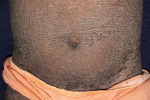 Epidermolytic ichthyosis
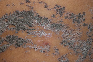 Epidermolytic nevus