Answer to Image of the Month April 2014
Submitted by Rajalakshmi T, Isha Garg
Trichoepithelioma
The structure within the circle is the papillary mesenchymal body/germ and papilla structure
The images show a circumscribed neoplasm in the dermis arranged in a cribriform fashion and cell nests. These cells have scant cytoplasm and dark staining nuclei and show striking peripheral palisading. This is a characteristic feature of neoplasms showing follicular differentiation.
Infundibular differentiation is also seen, characterised by cystic spaces containing laminated keratin and a lining of squamous cells. Occasioanlly, signs of differentiation to inner/outer root sheath may be noted.
The papillary mesenchymal body/germ and papilla represents the attempted formation of follicular germinative structures by the tumor. This is an important feature which is not found in basal cell carcinomas. However, this findings may be attenuated or absent in a desmoplastic trichoepithelioma, making the distinction between the two difficult.
In such an instance, the CD10 immunostain adds value by highlighting the mesenchymal component within this structure in case of trichoepithelioma, whereas the tumor cells are negative. In contrast, CD10 stains the neoplastic cells in BCC. Other useful markers include androgen receptor and cytokeratin 20.
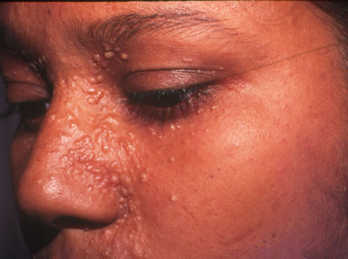 Trichoepitheliomas in the nasolabial folds and adjacent skin
(Image courtesy: M Ramam)
Clinically, solitary trichoepitheliomas are non-descript. Multiple trichoepitheliomas, however, have a recognisable appearance with hemispherical, yellowish papules that have a predilection for the nasolabial folds.