Answer to Image of the Month September 2012
Epidermodysplasia verruciformis
There are many enlarged keratinocytes predominantly in the upper epidermis. These keratinocytes have abundant bluish cytoplasm and large nuclei with clear nucleoplasm.
Verruca plana, on the other hand, show vacuolated keratinocytes in the upper epidermis and lack the cytoplasmic and nuclear changes seen here.
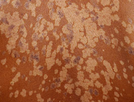 Multiple hypopigmented coalescing macules with interspersed pigmented macules and thin keratoses
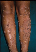 This patient had unusually large warts. Commonly, plane warts or small verrucae are seen.
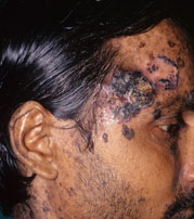 Basal cell carcinomas on sun-exposed skin. Note the small adjacent warts.
Clinically, there are multiple hypopigmented macules and thin plaques that superficially resemble pityriasis versicolor. In addition, there are warts of varying sizes and thickness. Squamous and basal cell carcinomas may supervene in some patients.
Uncommonly, the histopathological changes of epidermodysplasia verruciformis may be seen in biopsies from ordinary warts.