Answer to Image of the Month October 2013
Submitted by Meenakshi Batrani, Asha Kubba
Langerhans cell histiocytosis
The biopsy is from the back of a three month old female child who presented with papular rash involving trunk, flexures and scalp since the age of one month.
Sections show a band like upper dermal infiltrate obscuring the dermo-epidermal junction and infiltrating the epidermis. The infiltrate is composed of uniform large cells having abundant pale eosinophilic cytoplasm with central to eccentric reniform vesicular nuclei and admixed eosinophilis.
Immunohistochemisry showed positive staining of histiocytic cells with S-100 and CD1a (see image below) which is diagnostic for Langerhans cell histiocytosis. Non Langerhans cell histiocytoses are negative for S-100 and CD1a whereas the histiocytes in Rosai-Dorfman are positive for S-100 and negative for CD1a.
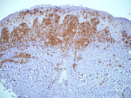 CD1a staining reveals that most of the histiocytes are Langerhans cells
A clinical image of a different patient appears below for illustration
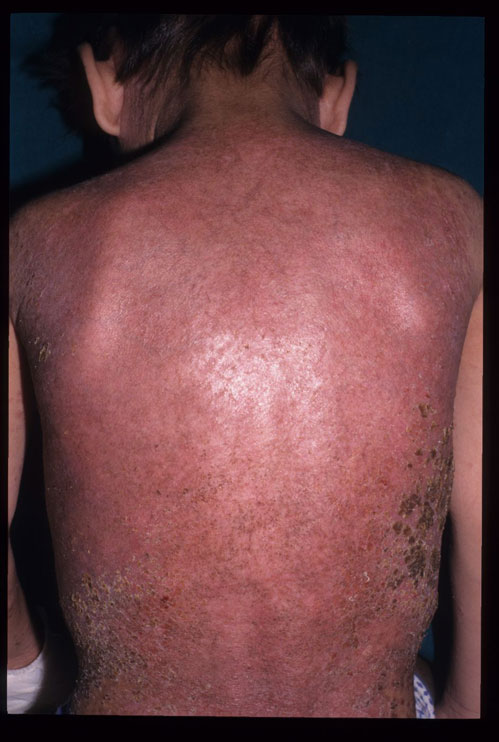 Scaly eruption on the back of a child with Langerhans cell histiocytosis (Image courtesy M Ramam)