Answer to Image of the Month June 2013
Submitted by I S Reddy, G Swarnalata
Herpetic blister
The image shows a intraepidermal bulla. The epidermal cells in the roof
of the bulla show ballooning and acantholysis. The bulla cavity shows
numerous acantholytic cells. Some of the acantholytic cells show
homogenous, eosinophilic cytoplasm. Basal cells are destroyed and the floor of the bulla shows naked dermal papillae. The inset shows numerous
multinucleate giant cells with eosinophilic intranuclear inclusion
bodies. This histological appearance is common to herpes simplex,
varicella and herpes zoster infections.
The image was of a biopsy from a 55-year-old male on a prolonged
course of thalidomide for erythema nodosum leprosum. The patient
presented with acute onset fever, generalised, polymorphic,
papulovesicular and pustular lesions and shortness of breath. Tzanck
smear showed numerous acantholytic cells and multinucleate giant
cells. Disseminated varicella was diagnosed and patient was started on
intravenous acyclovir and other symptomatic treatment. Patient was
intubated because of hypoxia and subsequently developed hypotension
and disseminated intravascular coagulation and expired 3 days after admission.
An example of a similar eruption in a different patient appears below.
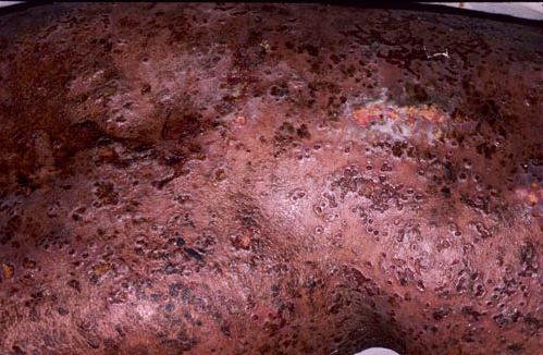 Disseminated varicella in a patient with pemphigus on immunosuppressive therapy (Image courtesy M Ramam)