Answer to Image of the Month July 2014
Submitted by Mani Makhija
Panfolliculoma
Panfolliculoma was a name coined by Dr A Bernard Ackerman in 1996 for a benign tumour of follicular differentiation where, unlike a trichoblastoma or pilomatricoma, the tumour shows differentiation towards all components of the normal hair follicle with its investing connective tissue and hence "panfolliculoma."
A trichoblastoma would show differentiation towards hair follicular germ with its papillary mesenchymal bodies; while a pilomatricoma would show only matrical differentiation evidenced by the ghost cells.
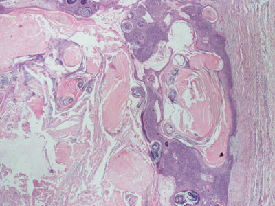 Fig 1: Sharply circumscribed border of tumour
Despite variation in size ranging from small to large (more than 1 cm), panfolliculoma has a benign silhouette, symmetry, sharp circumscription (seen in Figure 1) and forms stromal clefts as if it may be enucleated.
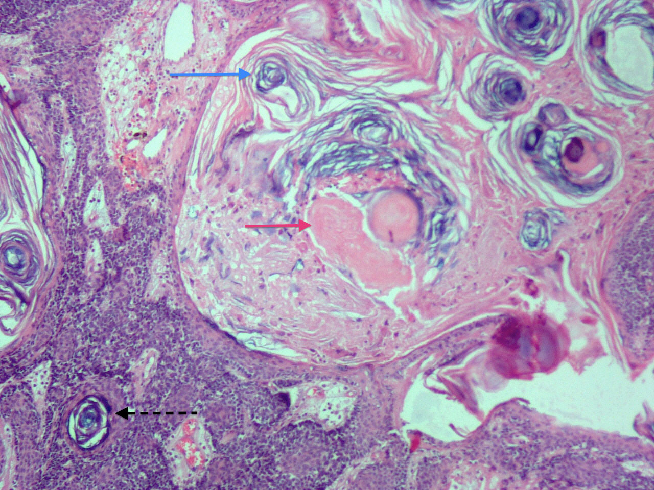 Fig 2 Blue and pink keratin (click to enlarge image)
 Fig 3 Higher power showing ghost cells (click to enlarge image)
The neoplasm may be solid, solid and cystic or predominantly cystic, but what is most striking on low power scanner view is a cystic area where lamellar keratin (figure 2 and 3 - blue arrow) is surrounded by blue germinative and matrical cell proliferation and shows areas of pink keratin which on higher magnification show matrical abortive hair formation i.e. "shadow" cells with "ghost" nuclei (Figure 2 and 3 red arrow). Unlike a pilomatricoma or matricoma, cysts in a panfolliculoma are also lined by infundibular epithelium with granular layer keratinising into lamellar whorled keratin like that in an infundibular (epidermoid or epidermal inclusion) cyst (figure 2 with dotted black arrow). Other areas may show bulbar, stem (with red trichohyaline granules), isthmic (with abrupt keratinisation) or infundibular differentiation and hence the name "pan"folliculoma.
The neoplasm may be considered as an expression of trichoblastoma with remarkably advanced follicular differentiation or a manifestation of matricoma, however, it has a distinctive and recognisable histology which warrants its identification as a separate distinct entity.