Answer to Image of the Month January 2013
Submitted by V Ramesh and Avninder Singh
Cylindroma
The histopathology shows numerous closely set islands of epithelial cells in the dermis separated by hyaline sheaths and narrow bands of collagen.These islands seem to fit together like pieces of a jigsaw puzzle. Two types of cells constitute the islands: cells with small dark staining nuclei at the periphery and cells with light staining nuclei towards the centre. The hyaline sheath is variable in thickness and hyaline droplets are present in some islands. Tubular lamina with and without amorphous material may be seen which at times may be numerous.
Clinically, cylindroma is an appendageal tumour, firm and pink-coloured, often solitary and predominantly presenting on the scalp and face. When multiple lesions occur on the scalp they are picturesquely referred to as turban tumours. The cell of origin of this tumour is unclear. The tumours show overlapping signs exhibiting apocrine, eccrine, secretory and ductal features. Treatment is by surgical excision.
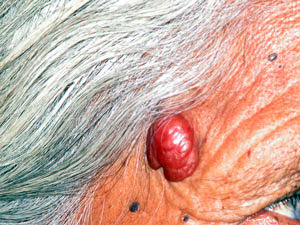 Solitary cylindroma on forehead