Answer to Image of the Month February 2015
Submitted by Meenakshi Batrani and Asha Kubba
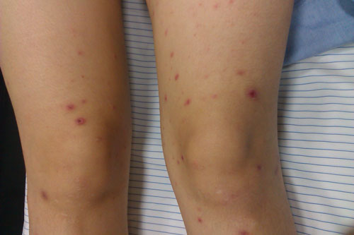
Papulonecrotic Tuberculid
The biopsy is from a papulo-necrotic lesion on the right buttock of a 21-year old woman who presented with recurrent crops of erythematous papulo-necrotic lesions on extensor surface of arms, thighs, legs and on buttocks. Lesions regressed leaving behind depigmented varioliform scars. The clinical differential diagnoses were perforating folliculitis versus pityriasis lichenoides et varioliformis acuta (PLEVA).
Histopathology showed a wedge shaped zone of necrosis. There was an infiltrate of lymphocytes, histiocytes and few giant cells forming occasional ill-defined granulomas at the periphery. There was no vasculitis. Mantoux test, done subsequent to the biopsy report, was highly reactive.
The typical lesion of papulo-necrotic tuberculid usually show a zone of necrosis. Surrounding granulomatous infiltrate and vasculitis are variably seen. The closest histopathological differential diagnoses are PLEVA and lymphomatoid papulosis.